Amoebiasis of bees

Amoebiasis is an invasive disease of the bee family, accompanied by the defeat of the Malpighian vessels of adult bees.
The causative agent of the disease is amoeba – Malpighamoeba mel-lificae.
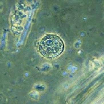
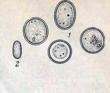
The parasite is a changing shape of the body, consisting of a nucleus and protoplasm. The amoeba nucleus strongly refracts light, and protoplasm reveals a clear differentiation into ecto – and endoplasm. Amoeba outside the body of the bee is preserved in the form of a cyst, which is a slightly oval or spherical-shaped body measuring 6-7 microns, covered with a smooth, dense, two-contour, hard-to-paint coat. Protoplasm, occupying the entire cyst space, strongly refracts light.

Fig. 23. Cysts (1) amoebae and spores (2) and form nostril stands.
It contains the nucleus, and in the nucleus there is a large nucleolus, which occupies almost all the nucleus. The cyst, when ingested with food or water into the body of a bee, turns into a vegetative form; The latter is introduced into the Malpighian vessels, where it develops.
Amoeba moves with pseudopodia – pseudopods, which are characterized by sharp points and annular bends.
In malpighian vessels, the amoeba densely sucks to the surface layer of epithelial cells, penetrates its pseudopodia between cells and extracts necessary nutrients from them.
Under unfavorable conditions of development, for example, with a lack of food, a decrease in temperature, amoeba, stop reproduction and form persistent cysts.
Before the formation of cysts, the nucleus of the amoeba acquires a dense structure, protoplasm, freed from excess water, concentrated and covered with a shell.
In the form of a cyst, an amoeba can remain for a long time even under adverse environmental conditions. Especially good cysts suffer drying.
Once in the body
Adult bees are susceptible to amoebiasis. Artificial infection is possible with feeding in sugar syrup the excrements of sick bees (containing cysts). Cyst formation in malpighian vessels occurs in 24-28 days.
Epizootic data. The source of the invasion are sick bees. Cysts of amoeba from Malpighian vessels are excreted together with excreta through the intestine into the external environment, contaminating food, nesting honeycombs, drinking bowls. In the future, through the contaminated feed, objects and water, healthy bees become infected.
The course and symptoms of the disease. Amebiasis often occurs as a complication of nosematosis and coincides with the course of the latter. During winter, amoebiasis is almost absent (1%), in March and April it increases rapidly (14%), in May it reaches its highest level (33%), and since June it has been noticeably decreasing.
Amebiasis usually occurs as a secondary invasion of nosematosis. Proceeding from the fact that the spread of amoebiasis is promoted by the same factors as in nosematosis, it must be assumed that the reasons that contribute to the development of nosematosis simultaneously contribute to the development of amoebiasis.
With a double invasion – nosematosis and amebiasis – the bees quickly become weaker, they soon lose their ability to work and soon die.
The diagnosis is based on the microscopy of smears prepared from Malpighian blood vessels by rubbing them with a glass rod on a slide.
Prevention of amebiasis, as in nosematosis, is based on improving the conditions of care, feeding and keeping bees. Falling honey from the fall is replaced with flower honey or sugar syrup, in winter rooms lower the humidity by ventilation.
Control measures are the same as for nosematosis.
Treatment of amebiasis with fumagillin gives the same good results as with nosematosis.
Amoebiasis of bees
