Reproduction and development of bees
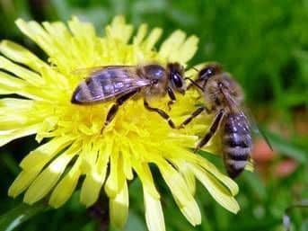
Growth and development of the bee family under favorable conditions of the environment (plenty of food, proper temperature, etc.) is provided by all three forms of individuals: the uterus, working bees and drones, but their role in this is not the same.
Uterus is the only fully developed female in the family. Its function is to postpone the eggs formed in its genital organs, from which all three groups of bee family can develop.
Working bees are also females, but their genitals are not fully developed, why they can not mate with drones and lay off fertilized eggs. However, working bees are perfectly adapted to perform other works necessary for the life of the family: brood rearing, nectar feeding and processing it into honey, collecting pollen, isolating wax and building, combs, nesting, etc.
Without taking a direct part in laying eggs, working bees are the nurse of the whole brood, the queens and drones and through the gland produced by the protein food (milk) affect the heredity (nature) of the next generations of the bee family.
Drones are males. Their role is in the fertilization of young queens.
Reproductive organs.
Breeding organs are well developed in the uterus and drone.
In the uterus, they fill a significant part of the abdomen and consist of two white, fairly large pear-shaped ovaries lying on the sides of the abdomen in the anterior half; in the fetal uterus, the ovaries reach a length of 7.5 mm, and in diameter – 4.5 mm. To the top, each ovary is thinned; The ovaries closely adjoin their curved vertices. Two egg tubes (one from each ovary), called paired oviducts, leave from the ovaries. These oviducts are connected to one common excretory duct – an unpaired oviduct. The lower extended part of this oviduct is called the vagina. Near the beginning of the general excretory duct, not far from the junction of paired oviducts, is a
The walls of the spermatheca are devoid of muscle. fly two writhing glands. These glands open into the duct of the spermatheca. The duct of the spermathecal can expand or be closed.
The spermathecal is used to preserve male sex cells-spermatozoa (seminal threads) obtained by the uterus when mating with the drones. In the unfertilized uterus, the spermatheca is filled with a transparent liquid, while in the fertilized one it is filled with millions of seminal threads, the purpose of which is to fertilize the eggs.
Ovaries consist of many egg tubules, which in both ovaries are from 200 to 360 or more, on average 300 pieces.
Eggs are produced in the egg tubes, and at the top each tube contains only the rudiments of eggs, which, as they mature, pass below; ripened eggs enter the paired oviduct, and thence into the unpaired or common excretory duct. The vagina opens outward under the sting.
In a worker’s bee, each ovary is most often composed of 4, but sometimes from a larger number to 20 poorly developed egg tubes and the spermatheca in its infancy.
Genital organs of the drone consist of two seminal glands, the so-called testes, having the form of beans and consisting of numerous – up to two hundred – convoluted seminiferous tubules. They produce sex cells.
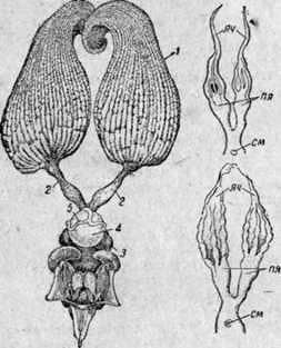
Fig. 19. Sexual organs of the uterus and worker bee: Left – the genital organs of the uterus: 1 – ovaries; 2 – paired oviducts; 3 – unpaired oviduct; 4-seminal receptacle; 5 – gland of the spermatheca; the genital organs of the working bee (from above) and the bee-work (from below) on the right: ovaries – ovaries; “I-paired oviducts, cm-spermatheca.
From each testicle departs one strongly curved tubule. These ducts end in significant extensions, the so-called seminal bladder, in which mature spermatozoa floating in the seminal fluid accumulate.
Seminal blisters open into the adnexal or mucous glands that produce a thick mucous fluid that is capable of quickly “congealing” when in contact with air. These glands are connected to one common long excretory duct, called the unpaired seed duct. Then this duct expands into a bean-shaped bladder (“bulb”), followed by a copulatory stump organ.
From the testes, the mature seed passes into the seminal blisters. From here, at the moment of pairing, the drones with the uterus move further and enter through the unpaired seed duct and the bean-shaped bladder. Following the seed, mucus enters there.
The sexual organ of the drone is arranged so that at the moment of mating, it is turned out by the inner side outward until the bean-shaped bubble and thus the bean-shaped bubble and its contents are pushed into the genitals of the uterus. Horny appendages of the copulatory organ do not enter the vagina of the uterus, but tightly close it, which is also facilitated by solidifying mucus. Thus, the seminal fluid can not flow out, but passes into the oviducts, where from 6-8 hours it is sucked into the spermatheca.
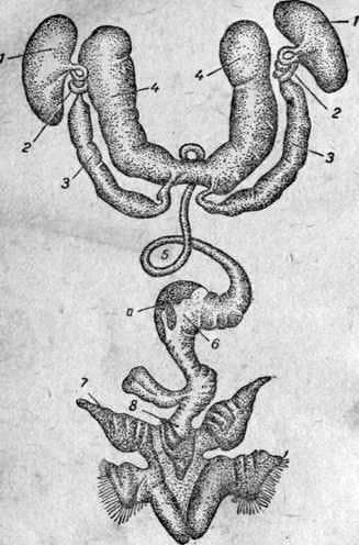
Fig. 20. Genital organs of the drone.
The twisted sexual organ of the drones breaks off when mating, and the drone dies.
If you press on the abdomen of a sexually mature drone, the sexual organ instantly turns out and is pushed out. The same happens when mating with the uterus.
If the air-bearing bags of the drone are densely filled with air, then it is easy to press these bags and abdominal muscles onto the genital organ, causing the latter to protrude and turn out. But in view of the fact that the drone only during flight can fill his air bags so tightly that the necessary pressure is exerted on the sexual organs, the fertilization of the uterus can occur only on the fly, hence outside the hive.
This is confirmed by experiments: a young uterus with circumcised or damaged wings remains barren.
Recently, scientists of the Institute of Apiculture found that the young womb mates not with a single drones, but with several – two, four, and in some cases even with a large number. At the same time, she can make several marriages from the hive, but can be covered in succession by several drones and for one flight. Since after each pairing a part of the genitalia of the drone or, as they say, the “trail” remains in the vagina of the uterus, each subsequent drone first releases the uterus from the trail left by the precursor drone, and then mates itself.
The uterus during the marital flight, lasting 15-20 minutes, mates until the seeds (seminal fluid) are paired with oviducts, and returns to the hive with a “train” of the last mating drones.
2-3 days after fertilization the uterus begins to lay fertilized eggs.
Fertilization of the egg.
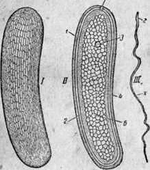
The egg has two shells, protecting it from damage. Outside the egg there is a fairly dense shell, and under it lies a very thin film, the so-called vitelline membrane. The upper dense shell on its entire surface is perforated with tubules. At one end of the egg there is a very small opening, which serves to penetrate the seed of the seed into the egg. The egg cell (egg) consists mainly of building material (yolk) for the body of the future larva.
 .
.
Fig. 21. Egg of the uterus (enlarged 35 times) and sperm of drone (increased 250 times). 1 – the appearance of the egg. II-egg in the section: 1 – egg shell; 2-yolk shell; 3 – the core; 4-zheltok; 5-hole, through which the sperm penetrates into the egg, the drone; b – protoplasm. 3 – spermatozoon: d – the head; x – tail, or flagellum.
In the seminal fluid of the drone, under a strong increase, one can see individual seed threads, or spermatozoa. The seed thread is the germ cell. One end of it is somewhat thickened, there is a core in it. This part is usually called the head of the seed thread. Spermatozoa have the ability to move.
For the fertilization of the egg, it is necessary that the seed thread penetrate into the egg through the above hole.
Eggs are fertilized when laying. It is believed that as soon as the egg enters the oviduct, the muscular apparatus surrounding the canal emerging from the receiver sucks in a part of the seminal fluid from the spermatheca.
The process of egg fertilization occurs in general so. Seed threads move to the opening of the egg and one of them enters the inside of the egg. Once in the egg, the sperm head begins to swell more and more and changes its shape. In it grows the central part – the core. Hour through 1-2 instead of the head of the seed thread, we find a male core, the same as the female one in the egg. Both nuclei gradually converge and after a few minutes merge.
After the fusion of the nucleus of the egg with the nucleus of the spermatozoon, the nucleation begins and the formation of new cells begins, from which the embryo is then formed.
Not all eggs are fertilized. From the fertilized eggs, females develop – working bees or uterus, and from unfertilized eggs – males, that is drones.
Unfertilized eggs have only maternal heredity. That is why, from the uterus, for example, of our local “breed”, fertilized by the yellow drones of the Italian “breed” of bees, her daughters – worker bees and queens – will be half-breeds, and her sons (drones) will be of the same local breed “, as the uterus.
The eggs laid by the mat are long and slightly curved corpuscles of white color. They reach 1.5 mm in length and about 0.5 mm in thickness.
The uterus lowers the egg, surrounded by mucus, to the bottom of the honeycomb cell. It sticks to the bottom of the cell with its lower end and remains in a standing, almost vertical position for the entire first day. On the second day the egg bends considerably; by the end of the third day it lies sideways, and then the larva emerges from it.
Development of working queens and drones of bees.
Bees develop the so-called complete transformation in their development, which consists in the following. Uterus lays eggs, from which larvae subsequently emerge. Then the larva turns into a pupa. Pupa does not eat. In the body of the pupa, all the organs are reconstructed, namely: the previous organs of the larval stage are fragmented into their constituent parts-cells and living matter of the non-cellular structure; new adult organs of an adult insect are created from this Material. Thus the pupa turns into a bee. Eggs of the larva and pupae are called brood. In this case, eggs and larvae before they are sealed are called open brood and larvae after sealing and pupae by a printed brood.
Fig. 22. Development of the bee (on days): A: 1-9 – open brood; from-11-printed brood 4-10-larva; 11 – pupa.
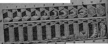 The larvae that have just emerged from the eggs are very small, milky white in color. They need careful care of the bees.
There are no air sacs in larvae, instead of them there are only large tracheal trunks.
The body of the larva consists of a head and 13 segments, of which the first three segments from the head are the chest and the rest are the abdomen.
External appendages absent. Only around the oral opening are observed oral parts in the form of 6 short outgrowths.
The eye has no larva; Instead of them there are only small protuberances.
On the sides of the larva, one can observe 10 respiratory holes called spiracles, or stigmata, which are branched throughout the body by breathing tubes (trachea).
All internal organs of the larva are clearly visible in Figure 23. The intestinal canal occupies most of the internal cavity. It consists of the anterior, middle and posterior intestines. Of these, the anterior and posterior are comparatively short and thin, and the middle is large, it occupies almost the entire internal cavity.
Above the intestine, the heart passes through the whole body in the form of a long thin tube lying under the skin of the back.
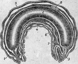
Fig. 23. Structure of the larva of the bee. Larva in longitudinal section (scheme):
1 – anterior gut; 2- medium bowel; 3- posterior gut;
4 – spinning iron; , 5 – heart; 6 – the nervous system; 7- Mal –
Piegive vessels; 8-ovary.
Between the heart and the middle intestine lie the paired parts of the genital organs.
The nervous system consists of 2 head and 11 abdominal knots and stretches under the intestine along the ventral side. The fatty body is up to 65% of the weight of the larva.
On the border between the middle and the back of the intestines, the urinary canals called the Malpighian vessels flow into the intestine.
The larva has two more spinning glands in the form of thin long vessels. They open on the lower lip and serve to curl the cocoon after the bees seal the larva in the cell.
All free places between the organs are colorless blood and fatty body.
The structure of the intestine of the larva has the feature that the junction of the middle and posterior intestines is closed by a thin septum, and therefore the contents of the midgut can not penetrate into the posterior. Because of this, all intestines, leftovers of food, perceived by the larva for 5-6 days of her life, are deposited in the midgut. They accumulate so much that this bowel is greatly inflated.
Only at the end of the larval life, shortly before the transition to the stage of the pupa, a thin septum between the middle and the hindgut bursts under the pressure of the garbage collected in the intestine, and the whole mass is pushed into the hindgut.
Feeding the brood. The process of embryo development in the egg at normal temperature (34-35 њ) lasts approximately 72 hours. When the temperature of the nest is lowered, the development slows down somewhat (sometimes reaching 80 hours), and at a slight increase accelerates (contracting to 68 hours).
By the end of the third day, the bees are poured food – milk – under the egg, lying on the bottom of the honeycomb cell, as a result of which its shell bursts and the formed small larva appears to be floating in the stern. If, for some reason, bees are not placed in a cell with an egg of liquid food (milk), the larval exit may be delayed for two days until the feed is given. This circumstance is sometimes used in the transport or transfer of eggs laid by breeding queens. With such transportation take measures to ensure that the air around the honeycomb with eggs was sufficiently wet, otherwise the eggs can dry. For this, the honeycomb is wrapped in a damp towel or in paper, which from time to time must be moistened.
Larvae are abundantly supplied with food. In the first days, the weight of the feed is 3-4 times the weight of the larvae, which lie, bent in a semicircle at the bottom of the cells, and slowly moving around in a circle, eagerly eat food.
Bee and drone larvae at an early age receive a different feed than in the later. Particularly different in composition is the feed given to the uterine larvae.
Although the uterus and worker bees are derived from identical fertilized eggs, their larvae, fed by unequal food, develop in different ways, resulting in either a fully developed female (uterus) or a not fully sexually developed female, that is, a working bee. This particularly vividly confirms the teaching of Michurin biology about the influence of the environment on the formation of the organism.
The newly hatched larvae are fed with so-called milk.
This milk is very nutritious, since it consists of protein compounds, fat and carbohydrates, and also contains vitamins and minerals and has no indigestible substances. According to the research, milk contains: protein 30.62%, sugar 15.05%, fat 15.22%, as well as other substances, so it is a fully-fledged food.
Bee larvae feed on milk only during the first three days. From the end of the third or from the beginning of the fourth day, the bees-nurses feed them with coarser food, the so-called gruel, which consists mainly of flower pollen (or pergus), honey and milk. The midgut of the larvae begins to shine through the color that the pergus has eaten.
Trout larvae are fed with milk a little longer, since often on the fourth day there are no pollen in their intestines.
As for the uterine larvae, they are fed throughout the entire larval life only with milk, that is, the most nutritious and digestible protein food, which makes the uterus develop rapidly.
Royal larvae are given so much food that they do not eat it completely. After the exit of the uterus, the remainder of the feed can be found in the mother liquor.
Larvae of bees and queens in the first 24 hours after leaving the egg have basically the same structure. On the second day, the genital organs of the larva of the worker’s bee seem to stop in their development. That’s why the queens should be removed from the larvae of the youngest age.
By changing the food, you can get not only dwarf or very large queens and working bees, but also transitional forms, that is, creatures in which the signs of the uterus and the worker’s bee are combined. As an exception, one can observe the development of a bee in a drone cell, and a drone in a uterine or bee. The development of the drone in the uterine cell does not reach the end, since it usually dies in the stage of the larva or pupa. The sealed mattock with the drone differs from the uterus containing its abnormal length and constriction in the middle like a cocoon of a silkworm.
Molting larvae and printing cells. Thanks to nutritious and abundant food, the larvae grow rapidly. From small, hardly appreciable they in some days turn to thick, hardly containing in the cells. The larvae lie in a cell, curled in an annular manner, and touching the sides of the cell with their backs. Most quickly they grow on the third and fourth day. For 6 days the larva increases in weight 1300 times. With such rapid growth, the larva fades four times in a short period of its existence. Discarded by the larva, the peel remains at the bottom of the cell.
At the end of the fifth day, the larva of the working bee becomes so large that it no longer fits on the bottom of the cell. On the sixth day fills the entire cell and begins to stretch along the cell, head outward. At this time, the bees are sealed with a lid, which is made not with a single wax, but with an admixture of perga, so that the lid is porous and lets in air.
In the sealed cell, the larva straightens, attaches to its cap the filaments of its semi-cocoon. With these threads, the larva is used in the same way that many insects do it before pupation. The threads needed for this purpose are produced in so-called spinning glands that open on the lower lip.
The larva of a silky spider web fills the sheath, the walls and the bottom of the cell, forming a cocoon. In the formation of the cocoon, part of the larval skin also plays a part.
Pupa and turning into a bee. After the ordering, the so-called pre-pupal stage follows; Inside the body of the larva, very complex changes occur: most of the former organs seem to dissolve, and instead of them new organs of a different structure are created. This state is called turning into a pupa. Finally, the larva fades. This is the 5th molt, or molting on a chrysalis. The discarded skin is the inner lining of the cell and, together with the cocoon, separates the chrysalis from the excrement discarded by the larva.
After the moult, the pupa is reminiscent of a swaddled bee.
Pupa is at first soft and has a white color, and then begins to gradually darken. The skin hardens and becomes covered with hairs. Finally the pupa ripens completely. Then she drops her skin. This is the last molt, that is molting on the bee, after which the young bee is fully formed. “Shirt” pupa remains lying in the depth of the cell.
The uterine larva curls the cocoon under the lid and not completely from the sides. The bottom is left without a cloth cloth.
In the event that bees destroy the maturing queens, they always gnaw the mother on the side where the cocoon is gone.
Approximately one day before the working bee or uterus leaves, bees gnaw wax from the lid and expose the cocoon. This facilitates the output of the bee and the uterus. The uterus and drones gnaw the lid on the edge in such a way that it usually disappears in the form of a mug. Working bee gnaws through the middle of the lid.
The uterus is vigorous and quite strong, and the workers and drones are much weaker.
The time required for the development of the uterus, bees and drone. For the transformation of the uterus from the testicle into an adult insect, an average of 16 days is required, namely: the larva is born from the egg after 3 days; in unsealed form, the larva remains for 5 days and in sealed form until the cell leaves the mature uterus – 8 days. The better the larvae of queens are raised, the faster their development and the better quality they are. Under very good conditions, the development of the uterus can last only 14 days.
It is generally believed that 20 to 21 days are required for the withdrawal of the bee, namely: the larva is born from the egg after 3 days; in unsealed form, the larva is about 6 days and in sealed form until the young bee leaves the cell about 11-12 days, and only -20 – 21 days.
It takes about 24 days to remove the drone, namely: the larva is born from the egg after 3 days; in unsealed form, the larva remains about 7 days and in sealed form until the exit of the drone from cell 14, only 24 days.
Table 1
The average period of transformation of the uterus, worker bee and drone
Terms of transformation (in days) | |||
Periods of development | Uterus | Working bees | Drone |
The larva is born from the egg through an unsealed larva In sealed form: | 3 5 2 1 5 | 3 6th 2 1 2 7-8 | 3 7th 3 4 7 |
Total. | 16 | 20-21 | 24 |
The hatched bee is in the form of an adult. Its weight is approximately 100 g, body length 13-14 mm. In the future, it does not increase in size and in most of its organs there is no noticeable change.
Reproduction and development of bees
